
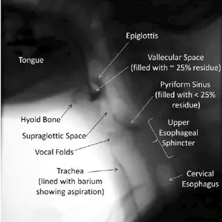
In radiology and the clinical setting, the Modified Barium Swallow Exam (MBSE) is commonly used to help diagnose aspiration in patients when food or liquid enter into the lungs and assess various medical conditions. It is a x-ray video fluoroscopic procedure that provides invaluable insights into the anatomy and function of the upper digestive tract. This essay delves into the anatomy, steps, diagnoses, and findings associated with the Modified Barium Swallow Exam. Stay tuned for a summary table as well as practice questions and answers at the end!
Anatomy of the Upper Digestive Tract:

The Modified Barium Swallow Exam (MBSE) is a radiologic procedure that provides detailed insights into the anatomy and function of the upper digestive tract during swallowing. Several anatomical landmarks and organs play a crucial role in this process. Let’s explore these anatomical structures:
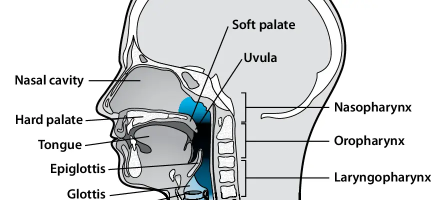
1. Oral Cavity: The oral cavity, also known as the mouth, is the initial entry point for food and liquids. It is bounded by the lips anteriorly, the cheeks laterally, the palate superiorly, and the tongue inferiorly. The oral phase of swallowing involves chewing, mixing food with saliva, and forming a bolus that can be easily swallowed.
2. Pharynx: The pharynx is a muscular tube that connects the oral cavity to the esophagus. It has three parts: the nasopharynx, the oropharynx, and the laryngopharynx. During swallowing, the pharynx plays a critical role in directing food and liquids away from the airway (trachea) and into the esophagus.
3. Epiglottis: The epiglottis is a flap-like structure located at the base of the tongue in the pharynx. During swallowing, it folds over the entrance to the trachea, preventing food and liquids from entering the airway and directing them toward the esophagus.
4. Larynx (Voice Box): The larynx houses the vocal cords and is situated just below the pharynx. It closes off during swallowing to protect the airway and prevent aspiration of food or liquids into the lungs.
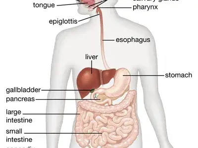
5. Esophagus: The esophagus is a muscular tube that connects the pharynx to the stomach. It propels food and liquids downward using coordinated contractions known as peristalsis. The lower esophageal sphincter (LES) separates the esophagus from the stomach, preventing the backflow of stomach contents.
6. Stomach: The stomach is a J-shaped organ located below the diaphragm. It receives food from the esophagus and breaks it down further through mechanical churning and the action of gastric juices.
7. UES (Upper Esophageal Sphincter) and LES (Lower Esophageal Sphincter): The UES is a muscular ring that separates the pharynx from the esophagus, helping to control the passage of food and liquids into the esophagus. The LES is located at the junction between the esophagus and stomach and prevents gastric contents from flowing back into the esophagus.
During the Modified Barium Swallow Exam, these anatomical landmarks and organs are closely observed using real-time X-ray imaging as the patient swallows various consistencies of barium-coated foods and liquids. The exam assesses the coordination, timing, and efficiency of the swallowing process, as well as the function of the various sphincters and structures involved.
By visualizing the movement of barium-coated materials through these anatomical structures, radiologists and healthcare professionals can identify any abnormalities, such as muscle weakness, structural issues, or neurological impairments that may affect the patient’s ability to swallow safely and efficiently. This information guides the diagnosis and treatment of conditions such as dysphagia, aspiration risk, and other swallowing-related disorders.
Steps of the Modified Barium Swallow Exam:
The Modified Barium Swallow Exam (MBSE), also known as a videofluoroscopic swallow study, is a diagnostic procedure that assesses the swallowing function and anatomy of the upper digestive tract using real-time X-ray imaging. Here are the steps involved in conducting an MBSE:
1. Patient Preparation: The patient is positioned in front of a fluoroscopy machine, which is a specialized X-ray imaging device that captures continuous images in real-time. The patient’s height and weight may be measured to determine appropriate barium doses for different consistencies.
2. Barium Preparation: Barium sulfate, a contrast medium visible on X-rays, is mixed with various consistencies of foods and liquids to mimic a patient’s typical diet. Different textures such as thin liquids, thickened liquids, pudding, and solid foods can be prepared, depending on the patient’s needs.
3. Initial Imaging: Before swallowing begins, a baseline X-ray image is taken to visualize the patient’s anatomy without any barium in the oral cavity or throat.
4. Oral Phase: The patient is instructed to swallow small amounts of the barium-coated foods and liquids in a controlled manner. The radiologist captures real-time X-ray images as the patient chews, forms a bolus, and propels it toward the back of the mouth in preparation for swallowing. This phase evaluates the oral control, lip seal, and formation of the bolus.
5. Pharyngeal Phase: As the patient swallows, the radiologist captures X-ray images of the bolus as it moves through the pharynx. This phase assesses the timing and coordination of various swallowing-related events, including the closure of the airway (epiglottis), movement of the vocal cords, and the initiation of peristalsis in the esophagus. The goal is to prevent aspiration of the bolus into the airway.
6. Esophageal Phase: The patient swallows a barium-coated liquid or solid, and its movement through the esophagus is monitored by the radiologist. The peristaltic waves that propel the bolus down the esophagus and the functioning of the lower esophageal sphincter (LES) are observed.
7. Additional Consistencies and Maneuvers: In some cases, different consistencies of barium-coated materials or specific maneuvers may be introduced to assess the patient’s swallowing ability in various scenarios. For example, a “chin tuck” maneuver may be used to observe changes in swallowing function when the patient tucks their chin to their chest during the swallow.
8. Observations and Analysis: Throughout the exam, the radiologist carefully observes the recorded images in real time, noting any abnormalities in swallowing coordination, timing, and anatomy. These observations are used to diagnose any issues with swallowing function and to identify potential causes of dysphagia or aspiration.
9. Report and Recommendations: After the exam is completed, the radiologist compiles their findings into a detailed report, which is shared with the referring physician or healthcare team. Based on the results of the MBSE, appropriate interventions or further diagnostic tests may be recommended to address any swallowing-related concerns.
In conclusion, the Modified Barium Swallow Exam is a comprehensive procedure that involves a series of steps to assess the swallowing function and anatomy of the upper digestive tract. By utilizing real-time X-ray imaging and barium-coated materials, this exam provides invaluable insights into the patient’s ability to swallow safely and efficiently, allowing for accurate diagnosis and tailored treatment planning.
Diagnosis and Findings:
The diagnosis and findings obtained from a Modified Barium Swallow Exam (MBSE) are crucial for identifying and understanding various swallowing-related issues and conditions. This diagnostic procedure provides detailed insights into the function and anatomy of the upper digestive tract during swallowing. Here are some common diagnoses and findings that can be identified through an MBSE:
1. Dysphagia: Dysphagia refers to difficulty in swallowing and can manifest as problems with initiating the swallow, moving food or liquids through the throat, or clearing the throat after swallowing. The MBSE can pinpoint the specific phase of swallowing that is causing the difficulty and help determine the underlying cause, such as muscle weakness, neurological disorders, or structural abnormalities.
2. Aspiration Risk: Aspiration occurs when food or liquid enters the airway (trachea) instead of passing through the esophagus, leading to potential lung infections like aspiration pneumonia. The MBSE can identify instances of aspiration and help determine the severity of the risk, guiding recommendations for safer swallowing strategies and dietary modifications.
3. Structural Abnormalities: The MBSE can detect structural abnormalities in the upper digestive tract, such as strictures (narrowing) of the esophagus, diverticula (outpouchings) in the pharynx or esophagus, or any other anatomical irregularities that might be contributing to swallowing difficulties.
4. Delayed or Incomplete Swallow Reflex: During the MBSE, the radiologist can observe whether the swallowing reflex is triggered in a timely manner and if the sequence of muscular contractions required for a successful swallow is coordinated properly. Delayed or incomplete reflexes can indicate underlying neurological issues or muscle dysfunction.
5. Gastroesophageal Reflux Disease (GERD): GERD occurs when stomach acid flows back into the esophagus, leading to symptoms like heartburn and regurgitation. The MBSE can provide insights into how reflux might be affecting swallowing function and if there’s a risk of aspiration due to GERD.
6. Impaired Upper and Lower Esophageal Sphincter Function: The exam can reveal dysfunction of the upper esophageal sphincter (UES) and lower esophageal sphincter (LES), which are muscular rings that control the passage of food into the esophagus and prevent reflux into the esophagus, respectively.
7. Effectiveness of Swallowing Maneuvers: The MBSE can assess the effectiveness of specific swallowing maneuvers, such as the “chin tuck,” which is often used to improve swallowing safety in individuals with dysphagia. Observations during the exam can indicate whether such maneuvers are beneficial for the patient.
8. Coordination of Airway Protection: The MBSE allows the radiologist to observe how well the airway is protected during swallowing to prevent aspiration. This includes the movement of the epiglottis and vocal cords, as well as the timing of breathing and swallowing coordination.
9. Evaluation of Diet Modifications: The exam can guide recommendations for dietary modifications, such as adjusting the thickness of liquids or altering the texture of foods to ensure safer and more efficient swallowing.
In summary, the diagnosis and findings from a Modified Barium Swallow Exam are essential for understanding the underlying causes of swallowing difficulties and aspiration risks. By assessing various aspects of swallowing function, anatomical structures, and coordination, the MBSE enables healthcare professionals to tailor treatment plans, provide targeted interventions, and enhance the overall quality of life for individuals with swallowing disorders.

Summary Table:
| Aspect | Description |
|---|---|
| Anatomy | Evaluates upper digestive tract anatomy, including oral cavity, pharynx, esophagus, larynx, epiglottis, and upper stomach. |
| Steps | 1. Patient preparation 2. Barium preparation 3. Initial imaging 4. Oral phase 5. Pharyngeal phase 6. Esophageal phase 7. Additional consistencies and maneuvers 8. Observations and analysis 9. Report and recommendations |
| Diagnosis | Identifies and diagnoses various swallowing-related issues: 1. Dysphagia 2. Aspiration risk 3. Structural abnormalities 4. Delayed or incomplete swallow reflex 5. GERD 6. Impaired sphincter function 7. Effectiveness of maneuvers 8. Coordination of airway protection 9. Evaluation of diet modifications |
| Findings | Provides insights into: – Swallowing coordination and timing – Airway protection during swallowing – Upper and lower esophageal sphincter function – Structural abnormalities – Effectiveness of swallowing maneuvers – Aspiration risk – Impact of GERD on swallowing – Recommendations for interventions and diet modifications |
Practice Multiple Choice Questions and Answers
Question 1: Which anatomical structure plays a critical role in protecting the airway during swallowing in the Modified Barium Swallow Exam?
A) Lungs
B) Esophagus
C) Epiglottis
D) Stomach
Question 2: What is the purpose of mixing barium with foods and liquids during the MBSE?
A) To enhance the taste of the foods and liquids.
B) To provide a contrast medium visible on X-rays.
C) To make the foods and liquids easier to swallow.
D) To improve digestion of the foods and liquids.
Question 3: Which phase of swallowing involves the movement of the bolus through the pharynx and into the esophagus?
A) Oral phase
B) Nasal phase
C) Pharyngeal phase
D) Gastric phase
Question 4: What is the primary function of the lower esophageal sphincter (LES)?
A) It controls the flow of air into the lungs.
B) It prevents food from entering the trachea.
C) It prevents reflux of stomach contents into the esophagus.
D) It aids in the formation of the bolus.
Question 5: During which step of the MBSE does the radiologist observe the initiation of peristalsis in the esophagus?
A) Oral phase
B) Pharyngeal phase
C) Esophageal phase
D) Additional consistencies and maneuvers
Question 6: Which condition is often associated with aspiration risk, where food or liquids enter the airway?
A) Parkinson’s disease
B) Hypertension
C) Osteoporosis
D) Type 2 diabetes
Question 7: What is the main goal of the “chin tuck” maneuver used during the MBSE?
A) To improve breathing coordination
B) To assess upper esophageal sphincter function
C) To enhance taste perception
D) To prevent aspiration during swallowing
Question 8: Which phase of swallowing evaluates the coordination of muscles involved in oral preparation and bolus formation?
A) Pharyngeal phase
B) Esophageal phase
C) Nasal phase
D) Oral phase
Question 9: What role does the epiglottis play during the MBSE?
A) It prevents reflux of stomach contents.
B) It controls the flow of air into the lungs.
C) It protects the airway during swallowing.
D) It aids in the movement of food through the esophagus.
Question 10: What aspect of swallowing function can be assessed by observing the movement of barium-coated materials through the upper digestive tract during the MBSE?
A) Heart rate
B) Blood pressure
C) Vision acuity
D) Coordination and timing
Answers and Explanations
Question 1: Answer: C) Epiglottis Explanation: The epiglottis is a flap-like structure that covers the entrance to the trachea during swallowing, preventing food and liquids from entering the airway.
Question 2: Answer: B) To provide a contrast medium visible on X-rays. Explanation: Barium is mixed with foods and liquids to make them visible on X-ray images, allowing the radiologist to observe the swallowing process in real time.
Question 3: Answer: C) Pharyngeal phase Explanation: The pharyngeal phase involves the movement of the bolus through the pharynx and into the esophagus, ensuring that it is directed away from the airway.
Question 4: Answer: C) It prevents reflux of stomach contents into the esophagus. Explanation: The lower esophageal sphincter (LES) prevents stomach acid and contents from flowing back into the esophagus, minimizing the risk of reflux.
Question 5: Answer: C) Esophageal phase Explanation: The esophageal phase assesses the movement of the bolus through the esophagus and evaluates peristaltic contractions that propel the bolus towards the stomach.
Question 6: Answer: A) Parkinson’s disease Explanation: Parkinson’s disease is often associated with muscle weakness and coordination issues, increasing the risk of aspiration during swallowing.
Question 7: Answer: D) To prevent aspiration during swallowing Explanation: The “chin tuck” maneuver helps prevent aspiration by altering the position of the head and facilitating safer passage of the bolus through the pharynx.
Question 8: Answer: D) Oral phase Explanation: The oral phase involves chewing, mixing food with saliva, and forming a bolus. It evaluates the coordination of muscles in the oral cavity.
Question 9: Answer: C) It protects the airway during swallowing. Explanation: The epiglottis covers the entrance to the trachea during swallowing, protecting the airway from potential aspiration.
Question 10: Answer: D) Coordination and timing Explanation: Observing the movement of barium-coated materials through the upper digestive tract helps assess the coordination and timing of various phases of swallowing.
Check out these popular articles 🙂
Circulatory System: Blood Flow Pathway Through the Heart
Ectoderm vs Endoderm vs Mesoderm
Circulatory System: Heart Structures and Functions
Ductus Arteriosus Vs Ductus Venosus Vs Foramen Ovale: Fetal Heart Circulation
Cardiac Arrhythmias: Definition, Types, Symptoms, and Prevention
Upper Vs Lower Respiratory System: Upper vs Lower Respiratory Tract Infections
Seven General Functions of the Respiratory System
Digestive System Anatomy: Diagram, Organs, Structures, and Functions
Kidney Embryology & Development: Easy Lesson
Autocrine vs Paracrine vs Endocrine: What are the Differences?
Their Eyes Were Watching God: Mule Symbol
Shoulder Abduction Muscles: Medical Anatomy and USMLE
Cell Membrane Dynamics: Flippase vs Floppase vs Scramblase
Cell Membrane Fluidity: Factors That Influence and Increase the Cell Membrane Fluidity
Psychology 101: Crowd Psychology and The Theory of Gustave Le Bon
Introduction to Evolution: Charles Darwin and Alfred Russel Wallace
An Overview of Shorthand: History and Types of Shorthand
Calculus: Two Important Theorems – The Squeeze Theorem and Intermediate Value Theorem
What is the Cori Cycle? Quick and Easy Explanation: MCAT and USMLE
Copyright © 2023 Moosmosis Organization: All Rights Reserved
All rights reserved. This essay first published on moosmosis.org or any portion thereof may not be reproduced or used in any manner whatsoever
without the express written permission of the publisher at moosmosis.org.

Please Like, Share, and Subscribe to our Email List at moosmosis.org, Facebook, Twitter, Youtube to support our open-access youth education initiatives! 🙂


Great essay!
LikeLiked by 2 people
Thank you so much!
LikeLiked by 1 person
💯
LikeLiked by 1 person
Thank you so much! 💕🙏
LikeLiked by 1 person