

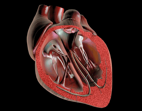
Heart Blood Flow Order
Blood flows through the heart in the following order: body –>inferior/superior vena cava –> right atrium –> tricuspid valve –> right ventricle –> pulmonary arteries –> lungs –> pulmonary veins –> left atrium –> mitral valve –> left ventricle –> aortic valve –> aorta –> body.

Vena Cava
The vena cava is the largest vein in the body that delivers oxygen-poor blood to the right atrium of the heart. The superior vena cava comes from the upper part of the body, including the brain and arms, while the inferior vena cava comes from the abdominal area and legs.
Atria
The atria are the top two chambers of the heart that receive incoming blood from the body. The right atrium receives deoxygenated blood through the superior and inferior vena cavas from the body and pumps it to the right ventricle through the tricuspid valve, which opens to allow the blood flow through and closes to prevent blood backing up the atrium. The left atrium receives oxygenated blood through the pulmonary veins from the lungs. It pumps the blood through the mitral valve to the left ventricle. Attached to the atria are the pouches called auricles that expand to allow the atria to include more blood volume.
Ventricles
The ventricles are the two lower chambers of the heart. The right ventricle receives oxygen-poor blood from the right atrium and pumps it through the pulmonic semilunar valve to the pulmonary artery and into the lungs to be filled with oxygen. On the other hand, the left ventricle receives oxygen-rich blood from the left atrium and pumps it through the aortic semilunar valve to the aorta to deliver the oxygen to the rest of the body.

Pulmonary Arteries
The pulmonary arteries deliver oxygen-poor blood from the right ventricle of the heart to the lungs while the pulmonary veins deliver oxygen-rich blood from the lungs to the left atrium of the heart.
Aorta
The aorta is the largest artery in the body that leads from the left ventricle of the heart to the rest of the body. It carries oxygen-rich blood to deliver to the body’s cells. As an artery, it contains thicker walls than veins because it has to withstand the tough pumping blood pressure of the heart.
Coronary Arteries
The coronary arteries are a set of arteries that branch off the aorta and are located on the heart. They carry oxygenated blood and nutrients to nourish the heart tissue cells. When the coronary arteries are clogged by excessive fatty tissue in cholesterol, it can lead to a lack of nutrients and oxygen for the heart, whose cells begin to perish, and this leads to coronary heart disease and/or myocardial infarction.
Pericardium
The pericardium is a tissue sac filled with fluid, which surrounds the heart to protect it, lubricate it, and absorb shock. It also attaches the heart to the chest cavity and separates the heart from the lungs in case of spreading infections.
Ductus Arteriosus
The ductus arteriosus is found in a fetal heart and provides a direct opening from the pulmonary artery to the aorta. Because the fetal lungs do not work and it receives oxygenated blood from the mother, blood can pass directly from the pulmonary artery (which leads to the lungs) into the aorta to be pumped to the rest of the body. The blood that leaks into the right ventricle after most of it passes from the right atrium to the left atrium through the foramen ovale is pumped into the pulmonary artery directly into the aorta through the ductus arteriosus. After birth, the ductus arteriosus is “pushed” closed by the lung’s need for the blood flow through the pulmonary arteries and into working lungs.

Foramen Ovale
In a fetus, the foramen ovale provides a shortcut for blood flow in the heart. Because the fetal lungs do not work, and the fetus receives its oxygen from the mother through the umbilical vein, the blood entering the right side of the heart already contains oxygen. Thus, it does not need to be pumped to the right ventricle to the pulmonary artery, lungs, and pulmonary vein. Instead, the foramen ovale allows an opening in which the blood can pass directly from the right atrium and into the left atrium. After birth, the infant’s foramen ovale will close and remain as a thin ligament between the atria.
Click and check out these awesome articles for more!:
Circulatory System: Blood Flow Pathway Through the Heart
Ductus Arteriosus Vs Ductus Venosus Vs Foramen Ovale: Fetal Heart Circulation
Copyright © 2021 Moosmosis Organization: All Rights Reserved
All rights reserved. This essay or any portion thereof
may not be reproduced or used in any manner whatsoever
without the express written permission of the publisher.

Works Cited
- Mori S, Tretter JT, Spicer DE, Bolender DL, Anderson RH. What is the real cardiac anatomy?. Clin Anat. 2019;32(3):288-309. doi:10.1002/ca.23340
- Miao JH, Makaryus AN. Anatomy, Thorax, Heart Veins. 2020 Jul 31. In: StatPearls [Internet]. Treasure Island (FL): StatPearls Publishing; 2020 Jan–. PMID: 31747193.
- Anderson RH, Wilcox BR. Understanding cardiac anatomy: the prerequisite for optimal cardiac surgery. Ann Thorac Surg. 1995;59(6):1366-1375. doi:10.1016/0003-4975(95)00195-q
- Iaizzo PA, Anderson RH, Hill AJ. The importance of human cardiac anatomy for translational research. J Cardiovasc Transl Res. 2013;6(2):105-106. doi:10.1007/s12265-012-9419-y
- Anderson RH, Yanni J, Boyett MR, Chandler NJ, Dobrzynski H. The anatomy of the cardiac conduction system. Clin Anat. 2009;22(1):99-113. doi:10.1002/ca.20700
- Miao KH, Guthmiller KB. Dextran. 2020 Oct 15. In: StatPearls [Internet]. Treasure Island (FL): StatPearls Publishing; 2020 Jan–. PMID: 32491563.
Share this:

Categories: Biology


95 replies »