
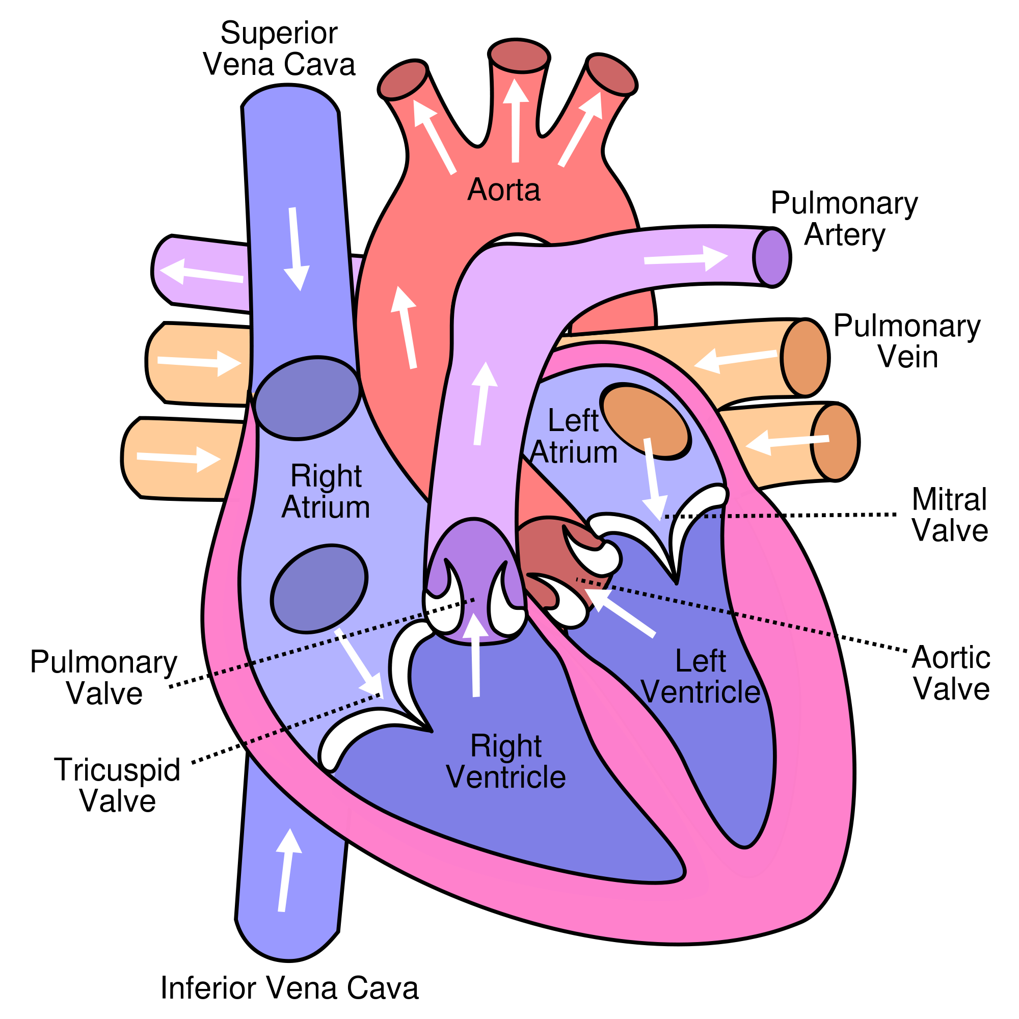
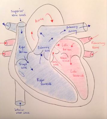
Pathway of Blood Through the Heart
In this educational lesson, we learn about the blood flow order through the human heart in 14 easy steps, from the superior and inferior vena cava to the atria and ventricles. Come also learn with us the heart’s anatomy, including where deoxygenated and oxygenated blood flow, in the superior vena cava, inferior vena cava, atrium, ventricle, aorta, pulmonary arteries, pulmonary veins, and coronary arteries.
Quick & Easy Video on Blood Flow Pathway Through the Heart
Blood Flow Order: Step by Step Animation Tutorial
To gain a visual step-by-step understanding, check out our quick and easy video on the blood flow pathway through the heart in less than 90 seconds. Please notice that blue represents deoxygenated blood, and red represents oxygenated blood.
14 Steps of Blood Flow Through the Heart
In summary from the video, in 14 steps, blood flows through the heart in the following order: 1) body –> 2) inferior/superior vena cava –> 3) right atrium –> 4) tricuspid valve –> 5) right ventricle –> 6) pulmonary arteries –> 7) lungs –> 8) pulmonary veins –> 9) left atrium –> 10) mitral or bicuspid valve –> 11) left ventricle –> 12) aortic valve –> 13) aorta –> 14) body.
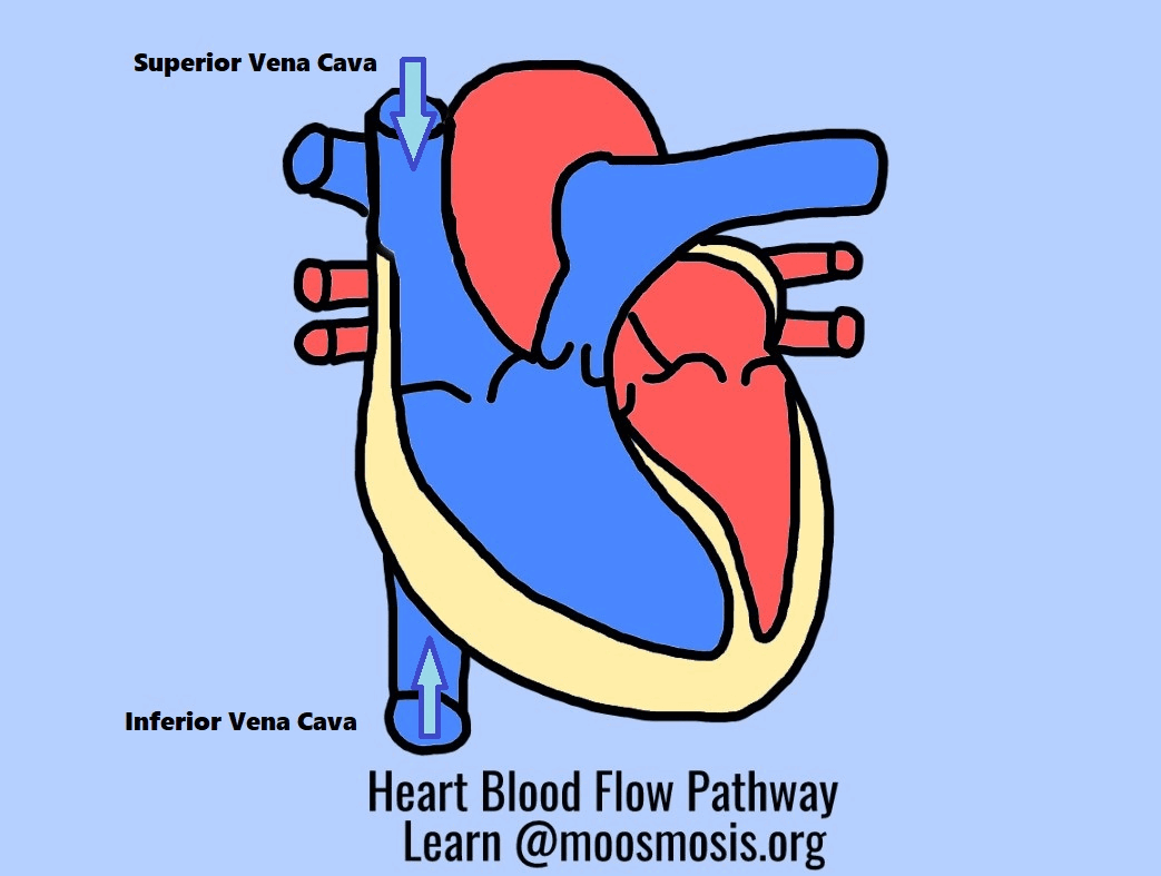

Superior Vena Cava & Inferior Vena Cava
The vena cava is the largest vein in the body that delivers oxygen-poor or deoxygenated blood to the right atrium of the heart. The superior vena cava comes from the upper part of the body, including the brain and arms, while the inferior vena cava comes from the abdominal area and legs.
Vena Cava and Inferior Vena Cava, respectively?
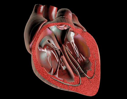
Left Atrium & Right Atrium
The atria are the top two chambers of the heart that receive incoming blood from the body. The right atrium receives deoxygenated blood through the superior and inferior vena cavas from the body and pumps it to the right ventricle through the tricuspid valve, which opens to allow the blood flow through and closes to prevent blood backing up the atrium. The left atrium receives oxygenated blood through the pulmonary veins from the lungs. It pumps the blood through the mitral valve to the left ventricle. Attached to the atria are the pouches called auricles that expand to allow the atria to include more blood volume. For fetal circulation, there is a special hole shunt between the left atrium and right atrium called the foramen ovale that diverts blood away from the lungs and goes directly to the rest of the fetus’s body.
passing through the right atrium into the right ventricle?
Left Ventricle & Right Ventricle
The ventricles are the two lower chambers of the heart. The right ventricle receives oxygen-poor blood from the right atrium and pumps it through the pulmonic semilunar valve to the pulmonary artery and into the lungs to be filled with oxygen. On the other hand, the left ventricle receives oxygen-rich blood from the left atrium and pumps it through the aortic semilunar valve to the aorta to deliver the oxygen to the rest of the body.
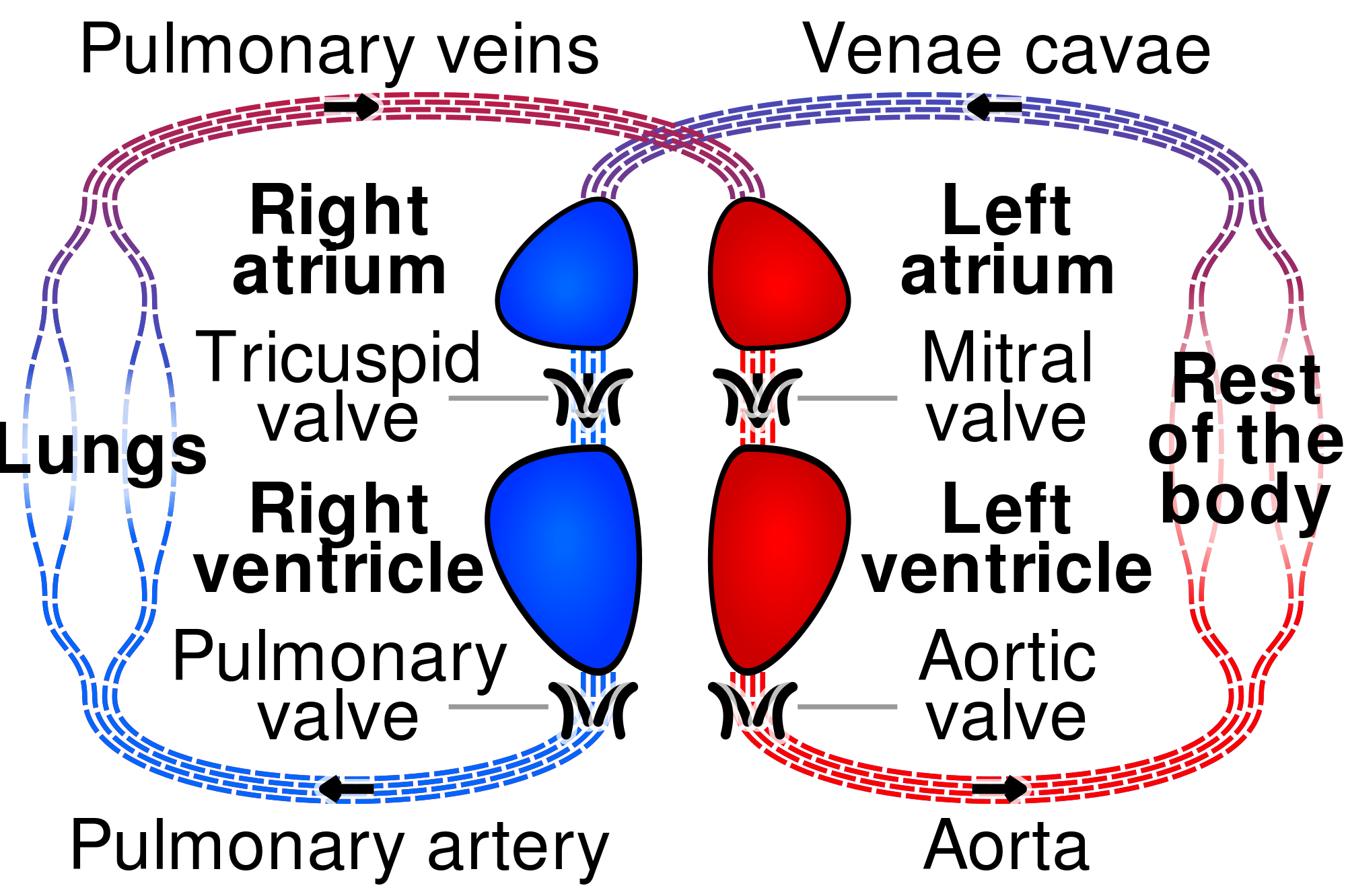
Pulmonary Arteries & Pulmonary Veins
The pulmonary arteries deliver oxygen-poor blood from the right ventricle of the heart to the lungs, while the pulmonary veins deliver oxygen-rich blood from the lungs to the left atrium of the heart. For fetal circulation, there is a special hole shunt called the ductus arteriosus that is between the pulmonary arteries and aorta to divert blood away from the fetus’s lungs. Learn more about how the ductus arteriosus works here, and why it’s there for fetuses.
delivers blood from the heart to the lungs?
Aorta & Coronary Arteries
The aorta is the largest artery in the body that leads from the left ventricle of the heart to the rest of the body. It carries oxygen-rich blood to deliver to the body’s cells. As an artery, it contains thicker walls than veins because it has to withstand the tough pumping blood pressure of the heart.
The coronary arteries are a set of arteries that branch off the aorta and are located on the heart. They carry oxygenated blood and nutrients to nourish the heart tissue cells. When the coronary arteries are clogged by excessive fatty tissue in cholesterol, it can lead to a lack of nutrients and oxygen for the heart, whose cells begin to perish, and this leads to a heart attack.
blood?
Final Test Your Knowledge! 😀
blood flow order through the heart is correct?
–> Inferior/Superior Vena Cava –> Right Atrium –> Right Ventricle
–>Pulmonary Arteries –> Lungs –> Pulmonary Veins –> Left
Atrium –> Left Ventricle –> Aorta –> Body
Ventricle –> Pulmonary Arteries –> Lungs –> Pulmonary Veins –>
Right Atrium –> Right Ventricle –> Aorta –> Body
Arteries –> Lungs –> Pulmonary Veins –> Right Atrium –> Right
Ventricle –> Inferior/Superior Vena Cava –> Body
Ventricle –> Pulmonary Veins –> Lungs –> Pulmonary Arteries –>
Left Atrium –> Left Ventricle –> Aorta –> Body
Summary: What are the 14 steps of blood flow through the heart?
Blood flows through the heart in the following order: 1) body –> 2) inferior/superior vena cava –> 3) right atrium –> 4) tricuspid valve –> 5) right ventricle –> 6) pulmonary arteries –> 7) lungs –> 8) pulmonary veins –> 9) left atrium –> 10) mitral or bicuspid valve –> 11) left ventricle –> 12) aortic valve –> 13) aorta –> 14) body.
Big thank you to our kind supporters! Please Like and Subscribe to our Email List at moosmosis.org, Facebook, Twitter, Youtube to support our open-access youth education initiatives! 🙂
Copyright © 2022 Moosmosis Organization: All Rights Reserved
All rights reserved. This essay or any portion thereof may not be reproduced or used in any manner whatsoever
without the express written permission of the publisher.

Please Like and Subscribe to our Email List at moosmosis.org, Facebook, Twitter, Youtube to support our open-access youth education initiatives! 🙂
*This article has been accepted into Moosmosis’s Journal of Global Health and Education . Accepted 2020. Published July 2020.
Works Cited
- Tubbs RS. The heart is simply a muscle. Clin Anat. 2016;29(3):267-268. doi:10.1002/ca.22704
- Miao JH, Makaryus AN. Anatomy, Thorax, Heart Veins. In: StatPearls. Treasure Island (FL): StatPearls Publishing; 2020.
- Anderson RH, Wilcox BR. Understanding cardiac anatomy: the prerequisite for optimal cardiac surgery. Ann Thorac Surg. 1995;59(6):1366-1375. doi:10.1016/0003-4975(95)00195-q
- Courchaine K, Rykiel G, Rugonyi S. Influence of blood flow on cardiac development. Prog Biophys Mol Biol. 2018;137:95-110. doi:10.1016/j.pbiomolbio.2018.05.005
- Efimov IR. Innovation in optical imaging: looking inside the heart. Heart Rhythm. 2007;4(7):925-926. doi:10.1016/j.hrthm.2007.04.006


Good and Simple learning
LikeLiked by 1 person
Thank you Vivek! Happy learning! 🙂
LikeLiked by 1 person
Wonderful diagrams and article on heart blood flow! Wholesome and helpful ❤️
LikeLiked by 1 person
Thank you so much Kay! Have a wonderful day ❤️
LikeLiked by 1 person
Good job done keep it up✌🏻
LikeLiked by 1 person
Thank you so much!!!
LikeLike
Thanks a lot of 👍
LikeLiked by 2 people
Excellent and extremely helpful! I was super confused about the order, this helped me understand the heart blood flow steps so much better, thanks!!
LikeLiked by 1 person
Thank you so much Jackie! Happy to help!
LikeLike
Very high level and comprehensive post! Thanks for visiting my blog. I can’t use the “like” buttons on your blog.
LikeLiked by 1 person
Thank you so much! You have a great blog too! Have a nice day!
LikeLike
Wonderful article on heart blood flow!
LikeLiked by 2 people
Thank you so much for your kind words, Susie!
LikeLiked by 1 person
This is so helpful! Made heart blood flow steps easy to understand, thanks!
LikeLiked by 1 person
You’re very welcome! So glad you found it helpful
LikeLike
Amazing! Excellent article on heart blood flow steps.
LikeLiked by 1 person
Thank you so much! Have an excellent day!! 😄
LikeLiked by 1 person
Excellent! So helpful
LikeLiked by 1 person
Thank you so much! We’re happy you found it helpful! ❤️❤️❤️
LikeLike
❤️❤️❤️
LikeLiked by 1 person
Thank you!! We love you too dear reader!! ❤️❤️❤️
LikeLike
Thanks for signing on to my blog – as an instructor in anatomy and physiology for medical students I think this blog is very helpful!
LikeLiked by 1 person
Thank you so much!!! ❤️ Your blog is very helpful and wonderful too! We hope you have a great day!
LikeLike