
In this lesson, we explore the nervous system and share notes as part of the study guide series. We will explore the awesome brain and nerves! Topics include the Early Types of Neural Cells, Structure and Function, Astrocytes vs Microglia vs Ependymal Cells vs Oligodendrocytes vs Schwann Cells.
Check out our popular nervous system notes.
Types of Neural Cells
- Two types of neural cells: neurons (aka nerve cells) and glia (aka neuroglia, glial cells).
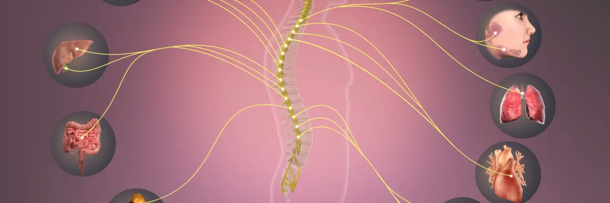
- Central nervous system = brain & spinal cord
- Peripheral nervous system = nerves coming out of spinal cord
- Nerves of PNS are made of neurons, glial cells, and other cells (which is why calling neurons “nerve cells” is problematic)
- Neurons are in both CNS and PNS, but different types of glial cells are just in one or the other.
- Most neurons and glia found in the CNSs are derived from neural stem cells, while most neurons and glia in the PNS are derived from neural crest cells.
- Both types arise early in embryo development, in the ectoderm.
- Most types of neurons and glia share structural features:
- soma — main body of cell that contains nucleus and most organelles
- long processes / projections that come out of the soma and vary in number, length, thickness, degree of branching, and terminal structures, as well as in their function.
- Function of neurons is to process and transmit information. Function of glia is to support the neurons in a variety of ways. (Even more glia than neurons.)
Neuron Structure
- Neurons consist of soma and projections called neurites, which are divided into dendrites & axons.
- Dendrites are short, branched projections often covered in small spines that increase their surface area and perform other functions
- Axons are longer and unbranched until it reaches its end. Might be short, or as long as 1 m or more.
- The end of the axon branches to create multiple axon terminals.
- Axon hillock = area where Axon leaves the soma
- Axon initial segment (trigger zone) = right after hillock, the first part of the axon projection
- Large axons are usually wrapped in a myelin sheath. Gaps that regularly interrupt the segments of myelin are called nodes of Ranvier.
- Axon terminals come close to the target cells (which might be another neuron, muscle cells, gland cells, or even capillaries (if releasing hormones to bloodstream), but don’t touch. This junction is called a synapse.
- Unipolar neurons — have soma and one projection (and axon)
- Inthe CNS — these start as neural stem cells, which can become any cell of nervous system, and then differentiate into (structurally similar) neuroblasts, which can only become neural cells.
- Neuroblasts then migrate away from other neural stem cells to the location their soma will be after development. They extend an axon, tipped with a growth cone, toward the target cell, which grows by following guidance cues in the environment until it reaches the target cell.
- In the PNS — both the original and migrating cells are neural crest cells instead of neural stem cells and neuroblasts
- Unipolar neurons are found in humans during fetal development, but not after.
- Bipolar neurons — have a soma, one axon, and one dendrite
- Multipolar neurons — have a soma, one axon, and multiple dendrites. This is most common type.
- Pseudounipolar neurons —have a soma, with short process coming out of soma that then divides into two long processes going in different directions. These are axons and the one bringing information in from the periphery is called peripheral axon, and axon bringing information into the CNS is called central axon.
- End of peripheral axon acts like dendrites do on other types of neurons. The part of the peripheral axon near the end is the initial segment / trigger zone, and the axon terminals are at the other end of the central axon.

Multipolar = motor neurons. motor neurons that conduct motor commands from the cortex to the spinal cord or from the spinal cord to the muscles
Pseudo-unipolar = sensory neurons. sensory neurons that receive sensory signals from sensory organs and send them via short axons to the central nervous system. Uni (one brain). Psuedo =PNS.
Bipolar neuron. Interneuron = . interneurons that interconnect various neurons within the brain or the spinal cord
Neuron Function
- Function of neurons is to process and transmit information.
- Most neurons have a resting membrane potential — a stable electrical charge difference across their membrane (more negative in the cell).
- This potential is how the neuron is able to be excited and to respond to input.
- Neurons receive excitatory or inhibitory input from other cells or from physical stimuli.
- Input info usually comes in through the dendrites, less often through soma or axon. The info from the input is transmitted to the axon with graded potentials — changes to membrane potential away from the resting potential. These are small in size, brief, in duration, and travel short distances.
- The size and duration of the graded potential is proportional to the size and duration of input.
- Summation = adding together of all the excitatory and inhibitory graded potentials at any moment in time. This summation occurs at the trigger zone, the initial axon segment, and is how neurons process information from their input.
- If the membrane potentials at the trigger zone crosses a threshold potential, information will be fired down the axon.
- Graded potentials are like a finger on the gun.. once pulled back a certain distance, information (bullet) will be fired down the axon.
- Action potential — a different change in membrane potential that allows information to be fired down the axon. These are usually large in size and brief in duration (they travel quickly), and can be conducted down the entire length of the axon, no matter how long
- Action potentials are usually the same size and duration for any particular of neuron. This is unlike graded potentials, whose size and duration depend on size and duration of input
- Action potentials travel faster down larger (bigger diameter) and more myelinated axons. When it reaches axon terminals, information (i.e. neurotransmitters) crosses the synapse gap to target cell.
- Neurotransmitters are released at axon terminal to bind to receptors on the target cell to maybe change that target cell’s behavior.
- Neurotransmitters are then removed from the synapse (via re-uptake channels) to reset system.
- Input information that was converted to size and duration of graded potentials is converted to the temporal pattern of firing of action potentials down the axon. This firing info is then converted to the amount and temporal pattern of neurotransmitter release at the synapse.
- Above is the general way neurons function, but there are multiple types of neurons:
- Afferent / sensory neuron— pseudounipolar that neuron brings info (about a stimulus) into CNS
- Efferent neurons — carry info away from CNS to PNS. Two main kinds of efferent neurons:
- motor neurons (aka somatomotor neurons) — control skeletal muscle
- autonomic neurons (aka visceromotor neurons) — control smooth muscle (e.g. around blood vessels), cardiac muscle, and gland cells.
- Autonomic neurons innervating the heart are responsible for releasing norepinephrine, a neurotransmitter of the sympathetic nervous system
- Most neurons of CNS aren’t like afferent/efferent, but are interneurons — they connect other neurons and form complex pathways for information to travel.
Astrocytes:
- Astrocytes are star shaped glial cells in the central nervous system (come from neural stem cells)
- Most common type of cell int the CNS
- Have lots of highly branched processes, at the end of which are end feet.
- They perform many many functions, possibly the greatest variety of functions, including the following:
(1) Form the scaffold for the whole CNS – give structural support to other cells in brain/spinal cord.
(2) Gliosis / astrogliiosis – involved in the repair and scarring process of the brain and spinal cord following traumatic injuries.
- If there’s an injury to the brain and/or spinal cord, astrocytes will divide and form more of themselves, migrate over to site of injury, and surround it.
- Their many projections then become hypertrophied and form a glial scar.
(3) Homeostasis of interstitial fluid — astrocytes take in or release necessary ions to keep environment for neural cells in homeostasis.
- Also release lactate (made from astrocyte glycogen) into interstitial fluid because neurons have very little internal energy in their cells and need that for energy.
(4) Blood-brain barrier – The end-feet of astrocytes are plastered all over blood vessels of CNS. These end-feet structures, along with components of the vessels themselves, form an effective barrier that prevents large molecules from leaving blood to enter CNS unless they want it
(5) Clears out synapses between neurons — Astrocytes place their end feet all over synapses and clear out neurotransmitters to reset the synapse for the next signal.
(6) Influence neurons and other glia through exchanging substances.
Most common cell type in the brain.
Astrocytes have structural, metabolic, regulatory, and repair functions, and are the most abundant cell in the brain.
Astrocytes are found in areas of brain scarring.
Astrocytes can supply lactate if the energy need arises.
Astrocytes are involved in strengthening the blood brain barrier, but do not monitor the interstitial fluid for foreign pathogens. This is the job of the microglia.

Microglia:
- Derived from mesoderm, instead of the ectoderm like all the other neural cells.
- Resting microglia have small soma and many highly branched processes heading out in every direction. In this state, they’re basically just monitoring the interstitial fluid looking for inflammation (from injury or infection). When they detect trouble, they convert to active microglia.
- The active microglia are just larger and sort of blob shapes. They act like macrophages and scavenge the CNS for plaques, damaged neurons, and infectious agents.
- Microglia are the resident macrophages of the of the brain and spinal cord, and thus act as the first and main form of active immune defense in the central nervous system. They do this by:
(1) Secreting cytotoxins — If a microglia finds a foreign cell it can secrete cytotoxic substances like reactive oxygen species that can kill a cell.
(2) Phagocytosis — After macrophages kill the bacteria, it becomes debris. Microglia eat up all kinds of debris, from foreign or from its own cell and break it all down. yum.
- Note: these processes don’t necessarily happen in order… They could phagocytose something and then secrete a cytotoxin.
(3) Antigen presentation — After consuming debris, microglia will take a tiny pieces of it and stick tjos antigen out on its surface for other cells (specifically those of the immune system) to see.
- Ex: Lymphocytes can then recognize the antigens, and potentially increase inflammation and/or make it more specific to whatever foreign cell the microglia has identified.
- Thus microglia is both activated by inflammation and it contributes to it.
(4) other cells of the immune system through exchange of substances.
Microglia arise from monocytes, and are a part of the immune system, which arises from the mesoderm. However, the majority of the nervous system arises from ectoderm (CNS) or neural crest cells (PNS).
Microglia are the macrophages, or phagocytes, of the central nervous system (CNS, or the brain). They will proliferate if there is an infection, such as bacterial meningitis.
Ependymal Cells:
- These cells line the CSF-filled ventricles in the brain and the central canal of the spinal cord.
- Derived from neural stem cells
- Ependymal cells are simple (one layer) columnar, cuboid epithelium-like cells.
- The side of the ependymal cell that faces the cerebral spinal fluid has many microvilli and cilia.
- Main functions:
(1) Form barrier between CSF and interstitial fluid of the tissue itself, though it’s a relatively leaky area (this leakiness if helpful because it means doctors can sample it).
(2) Secrete CSF — Specialized ependymal cells and capillaries form tufts in some of these brain ventricles. This is where CSF is secreted across ependymal cells to create cerebral spinal fluid.
Ependymal cells help form the barrier that holds in and produces CSF, cerebrospinal fluid. Ependymal cells not only help form the barrier that separates CSF from the rest of the body, but help secrete it as well.
The brain and spinal cord are cushioned by cerebrospinal fluid (CSF), which is kept separate from blood and lymph fluid.
Oligodendrocytes:
- Similar structure to astrocytes, but with fewer projections. Also in the central nervous system.
- main function: produce myelin sheath in the CNS
- Each oligodendrocyte has maybe a dozen projections that extend towards nearby axons of neurons. The structure at the end of these oligodendrocyte projections is myelin sheath.
- Each oligodendrocyte can produce myelin sheaths for multiple neurons (each projection makes one segment of the sheath, but they have multiple projections)
- The myelin sheath for a single neuron can come from multiple oligodendrocytes.
- Myelin sheath is made mostly of a lipid, the same substance that makes up fat, of course. It’s basically the cell membrane for an axon… sort of like the rubber coating on a wire.
- Oligodendrocytes also influence other glial cells.
Each nerve cell can have multiple myelin sheaths, which help speed conduction. Oligodendrocytes are myelin sheaths that wrap around nerve axons to help speed conduction.
Schwann Cells:
- Schwann cells are glia of the PNS, derived from neural crest cells. They come in different shapes.
- Nonmyelinating Schwann cells are fairly shapeless, but have troughs on their surface. Small diameter neurons can just sort of sit their axons in these troughs.
- These types of Schwann cells provide some support for PNS axons, but don’t myelinate them.
- The main function of normal Schwann cells is to produce the myelin sheath for PNS neurons.
- Not all peripheral neurons have a myelin sheath, but most of those with a larger diameter do. The structure and function of these myelin segments are the same in the PNS and in the CNS, but are produced by different cells.
- Schwann cells are also different from the oligodendrocytes of the CNS in that a single Schwann cell produces the myelin for a single segment of a single axon. They’re not myelinating multiple neurons like oligodendrocytes.
- Almost all the cell membrane of a Schwann cell is the myelin wrapped around an axon. It has just a little lump outside this wrapping, though, that contains the nucleus and cytoplasm for the Schwann cell
- Schwann cells also influence neurons, and vice versa, through exchange of various substances.
- Gangliosides are found on Schwann cells, the myelin sheath cells of the peripheral nervous system
- The majority of immune cells cannot cross the blood-brain barrier.
- Conduction potential velocity increases with increased axon diameter and myelination.
- Multiple sclerosis is an inflammatory disease, and is characterized by an increase in antibodies and a decrease in myelination.
Other:
- Tetanus is the maximum sustained contraction of skeletal muscle cells.
- Heart rate is controlled by the autonomic nervous system.
- Afferent nerve fibers bring signals back to the central nervous system.
- A local nerve block would be an anesthetic, as it would block sensation, but would not affect movement. Sometimes a nerve block is used to dull tooth pain when pain medication is contra-indicated.

Check out our popular nervous system articles!
Central Chemoreceptor vs Peripheral Chemoreceptor
Check out these popular articles 🙂
Circulatory System: Blood Flow Pathway Through the Heart
Ectoderm vs Endoderm vs Mesoderm
Circulatory System: Heart Structures and Functions
Ductus Arteriosus Vs Ductus Venosus Vs Foramen Ovale: Fetal Heart Circulation
Cardiac Arrhythmias: Definition, Types, Symptoms, and Prevention
Upper Vs Lower Respiratory System: Upper vs Lower Respiratory Tract Infections
Seven General Functions of the Respiratory System
Digestive System Anatomy: Diagram, Organs, Structures, and Functions
Kidney Embryology & Development: Easy Lesson
Psychology 101: Crowd Psychology and The Theory of Gustave Le Bon
Introduction to Evolution: Charles Darwin and Alfred Russel Wallace
Copyright © 2022 Moosmosis Organization: All Rights Reserved
All rights reserved. This essay first published on moosmosis.org or any portion thereof may not be reproduced or used in any manner whatsoever
without the express written permission of the publisher at moosmosis.org.

Please Like and Subscribe to our Email List at moosmosis.org, Facebook, Twitter, Youtube to support our open-access youth education initiatives! 🙂
Share this:

Categories: anatomy, Biology, cell biology, education, health, medicine, stem, technology


excellent article!
LikeLiked by 2 people